A newly developed nanocapillary system reveals that mouse embryo nuclei undergo a pronounced softening after the first cleavage division, a structural shift that proves essential for genomic activation and further development.
Nanocapillary Measurements Track Embryonic Nuclear Rigidity
Researchers working across Kyushu University and the National Institute of Genetics traced nuclear stiffness during mouse embryonic development. The collaborative group investigated the physical transitions occurring within the first 48 hours of mammalian life.
To capture these changes without destroying the cells, the laboratory of Yuta Shimamoto engineered a specialized micro-mechanical platform. This nanocapillary system that was developed by the Shimamoto lab allowed us to measure the stiffness of a cell’s nucleus, said Kei Miyamoto of Kyushu University.
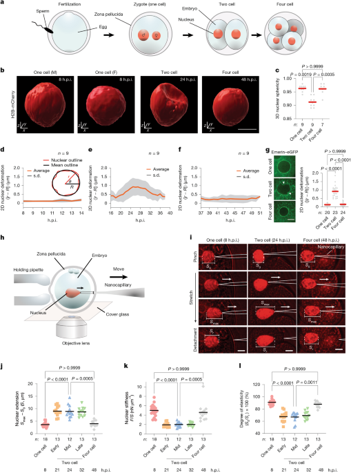
Measurements establish that the newly formed nucleus maintains high structural rigidity during the one-cell stage. Following the first cleavage division into the two-cell stage, the nuclear envelope becomes considerably softer, plastic, and visibly deformed. This pliancy is strictly transient; as the embryo divides further into the four-cell stage, the nuclei regain their baseline stiffness.
Lamin B1 Clearing Triggers Structural Compliance
Further biochemical analysis identified the mechanism driving this sudden pliability. Lamin B1, a protein that gives the nuclear envelope its structure, is cleared at the two-cell stage. Cells whose nuclei failed to soften at this critical juncture showed severely reduced developmental viability.
While we know that nuclear structure is important to life, we knew little about its role at the earliest stages of development, at the very beginning of life. We wanted to find out how the nucleus changes during the first few days after fertilization.
Kei Miyamoto, Kyushu University
The two-cell stage represents a critical window in embryonic life when major zygotic genome activation occurs. During this phase, approximately 2,000 genes become active almost simultaneously to kickstart embryonic progression.
Mechanical Compliance Drives Spatial Gene Organization
High-resolution imaging and locus-tracking experiments demonstrated that nuclear softening is functionally coupled to this transcriptional burst. In stiffened control nuclei, chromatin loci experienced constrained mobility that restricted developmental genes from shifting into active positions.
By contrast, the pliable envelope of the softened nucleus permitted active transcription sites to mobilize adjacent to the nuclear boundary. In embryos where researchers artificially suppressed nuclear softening, active DNA transcription near the periphery was markedly inhibited, demonstrating that mechanical compliance directly dictates transcriptional competence.
This was a very interesting finding. We knew that the two-cell stage is a critical time in embryonic development when a phenomenon called major zygotic genome activation, or ZGA, occurs. This is when over 2,000 genes become active almost simultaneously, kickstarting the development of the embryo.
Kei Miyamoto, Kyushu University
The experimental findings appeared in the journal Nature on November 11, 2026, establishing that mechanical deformation of the nucleus acts as an active functional regulator of genomic transcription rather than a passive consequence.
Applications for IVF and Cancer Research
Investigators see broad implications for both reproductive technology and oncology. Because the current study relied entirely on mouse models, the research team said that future work must determine whether transient nuclear softening governs early development in other mammalian species, including humans.
This is a great fundamental discovery for the field of developmental biology and holds significant potential in applied research.
Kei Miyamoto, Kyushu University
In the future, we hope this will lead to a better way of evaluating viable embryos in procedures such as IVF, and even to new tools for studying the physical properties of cancer cells.
Kei Miyamoto, Kyushu University