MIT Nanofibers Pave the Way for Customizable Cancer Vaccines
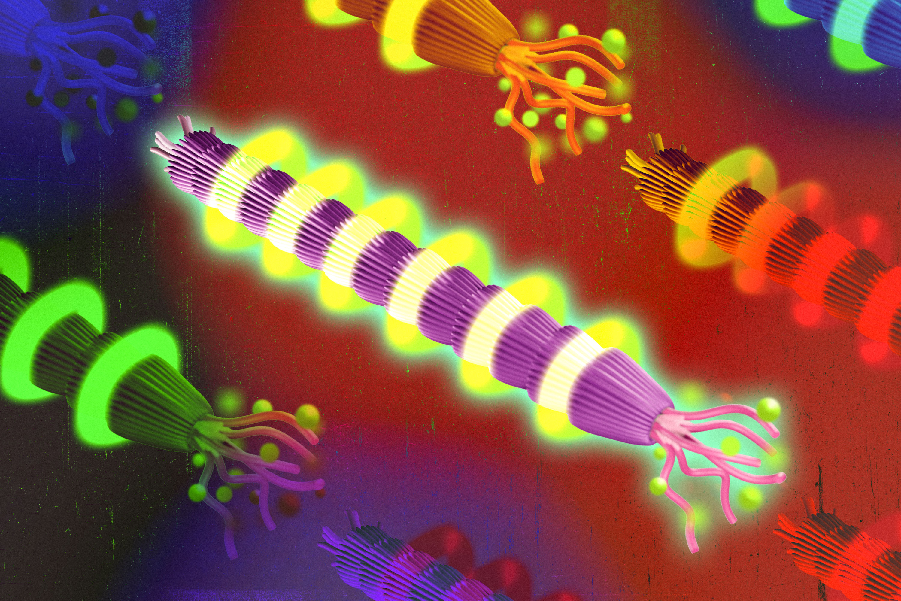
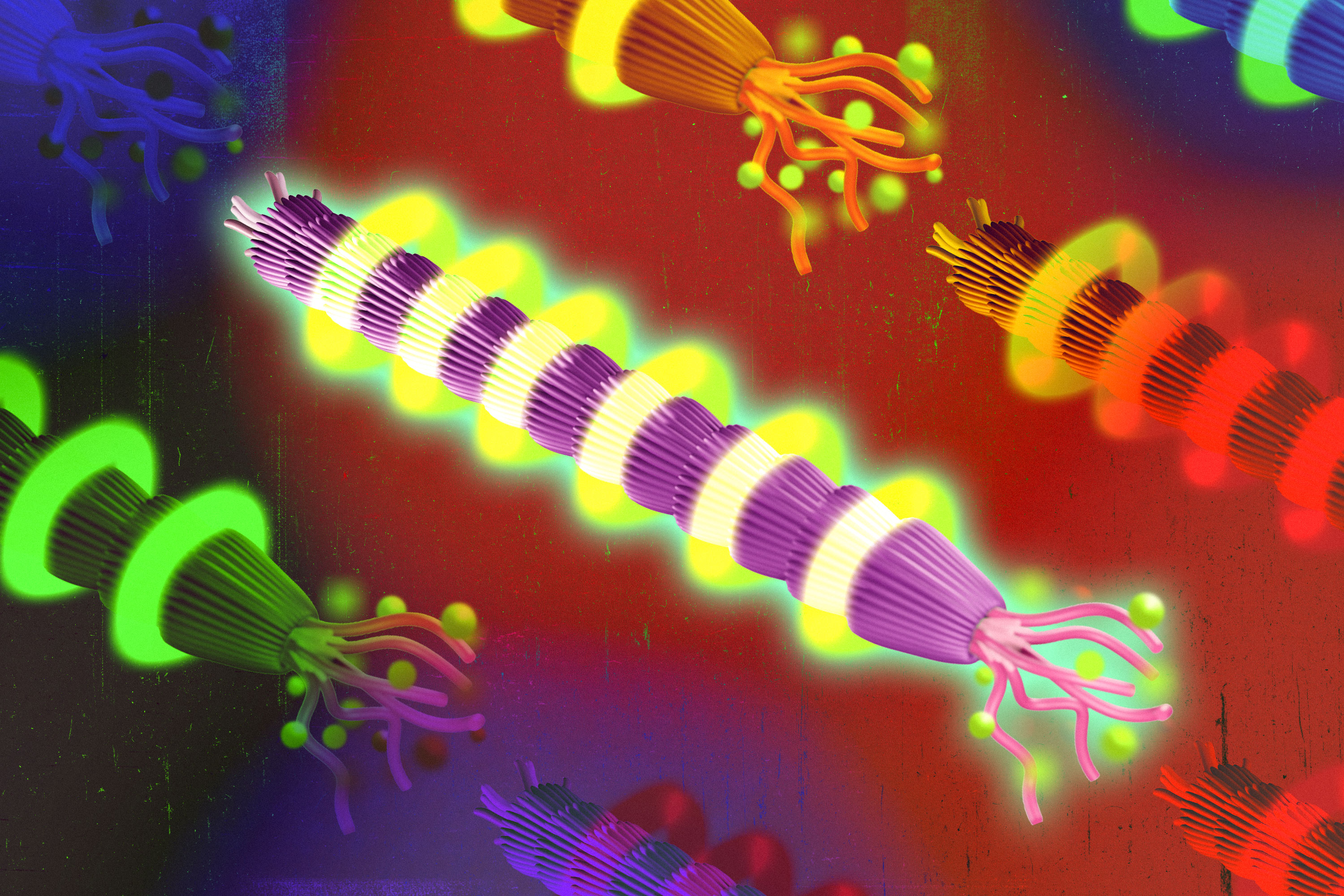
As reported in Advanced Healthcare Materials, the backbone of the system relies on a modified bacteriophage known as M13, which forms a nanofiber capable of displaying specific cancer antigens.
For more than two decades, the lab of Angela Belcher, the James Mason Crafts Professor of Biological Engineering and Materials Science and Engineering at MIT, has utilized the M13 bacteriophage to engineer materials for batteries, semiconductors, and solar cells. Recently, that decades-long toolkit has been adapted for medical therapeutics. By modifying the viral protein shell, researchers found they could precisely control the density of cancer proteins on the nanofiber.
Modifying Viral Coat Proteins to Display Target Antigens
The M13 virus consists of a single strand of DNA encased in a protein shell, or capsid. To construct the vaccine delivery platform, lead author and Koch Institute research scientist Shengnan Huang modified a specific coat protein called pVIII. By adding the gene for a target antigen directly into the virus’s DNA, the researchers caused the proteins to display the antigen on the end of the filament.
Controlling the exact dosage of the antigen proved critical for generating a strong immune response. Huang discovered that increasing the density of modified pVIII proteins was possible by adding one or two negatively charged amino acids to the protein sequence. Further fine-tuning was achieved by introducing a genetic mutation that slowed down the production of the antigen protein.
“The great thing about this is that it’s modular and we could switch out different antigens on demand, so you could design it to be patient-specific and target a known antigen for a particular kind of cancer,” Angela Belcher said.
In addition to carrying the antigen, the M13 phage naturally stimulates the body’s innate immune system. The single-stranded DNA of the virus activates a biological pathway called TLR9, allowing the particle to act as its own vaccine adjuvant. This eliminates the need to mix the vaccine with external adjuvants such as aluminum hydroxide, as the required components are entirely encoded within the genetic structure.
Testing Efficacy Against Tumors in Animal Models
To evaluate how antigen density impacts immune protection, the researchers tested particles expressing the model antigen SIINFEKL at densities ranging from 6 percent to 70 percent. Mice vaccinated with the 35 percent density particles generated more T cells and were much less likely to develop tumors when subsequently exposed to melanoma cells expressing the target antigen.
Alberto Danielli, an associate professor of molecular biology at the University of Bologna who was not involved in the research, noted that the study contributes a valuable mechanism to the translational potential of the M13 platform for cancer immunotherapeutics.