A 22-year-old man living in the United Kingdom experienced a sudden-onset left-sided weakness and numbness, leading to a rare medical diagnosis of an acute ischaemic stroke caused by cerebral neuroschistosomiasis, Cureus reported.
The patient, originally from Senegal, was brought to the hospital by ambulance after two days of developing difficulty speaking, headache, and poor balance. He had no significant medical history and no major cardiovascular risk factors. When questioned by clinicians, he reported a history of intermittent terminal haematuria that lasted for several days at a time over the preceding months, along with childhood memories of frequently swimming in freshwater lakes while resident in Senegal.
Neurological examinations revealed a left hemiparesis, sensory disturbance, dysarthria, and cerebellar signs in his left arm that were out of proportion to his limb weakness. His National Institutes of Health Stroke Scale score stood at 5 upon admission, dropping to 3 after 24 hours. While routine blood tests showed a normal white blood cell count and C-reactive protein, a full blood count revealed a marked peripheral eosinophilia of 1.4 × 10⁹/L, well above the standard reference range of 0 to 0.5 × 10⁹/L.
Neuroimaging Reveals Multifocal Intracranial Vasculopathy
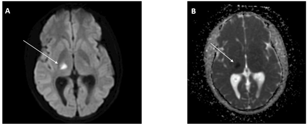
Non-contrast CT scans of the head showed a subtle hypodensity involving the right thalamus and the posterior limb of the internal capsule. Subsequent MRI confirmed an acute infarct in the right posterior thalamus with true diffusion restriction, showing no evidence of hemorrhagic transformation.
Doctors explored numerous potential causes for the extensive vascular changes. A comprehensive autoimmune and vasculitis screen—including PR3 and myeloperoxidase antibodies, antinuclear antibodies, complement levels, and an antiphospholipid screen—returned entirely negative results. Rheumatology specialists confirmed the clinical picture did not match a systemic vasculitis. However, the combination of peripheral eosinophilia, terminal haematuria, and a background of living in an endemic region sharply pointed toward a parasitic infection.
Urine microscopy ultimately confirmed the presence of Schistosoma haematobium ova, and serum schistosomal antibody testing via enzyme-linked immunosorbent assay returned positive. Additional imaging through CT intravenous urography displayed calcification of the bladder dome and an ectopic left kidney situated within the pelvis. Flexible cystoscopy further revealed a classical sandy patch lesion measuring 3 by 2 centimeters on the posterior bladder wall.
Oral Prednisolone Regimen Normalizes Eosinophil Count and Achieves Recovery
The patient received an oral prednisolone regimen tapering over six weeks, starting at 60 mg daily.
By six weeks post-treatment, repeat blood tests confirmed his eosinophil count had normalized to 0.5 × 10⁹/L, and post-treatment urine microscopy showed an absence of schistosomal ova. Over 12 months of clinical follow-up, the patient maintained a strong functional recovery with no recurrence of symptoms.