A Rare Surgical Success in the Face of a Complex Neonatal Diagnosis
In the high-stakes world of neonatal surgery, the combination of congenital pyloric atresia (CPA) and epidermolysis bullosa (EB) has long been viewed as a medical emergency with a historically grim prognosis. A recent report published in Cureus details the successful treatment of a five-day-old male neonate, offering a rare look at a benign, self-improving phenotype of this syndromic association. While past cases often resulted in high mortality rates due to systemic sepsis and severe skin fragility, this patient’s trajectory suggests that rapid surgical intervention and modern, atraumatic nursing care can lead to long-term survival.
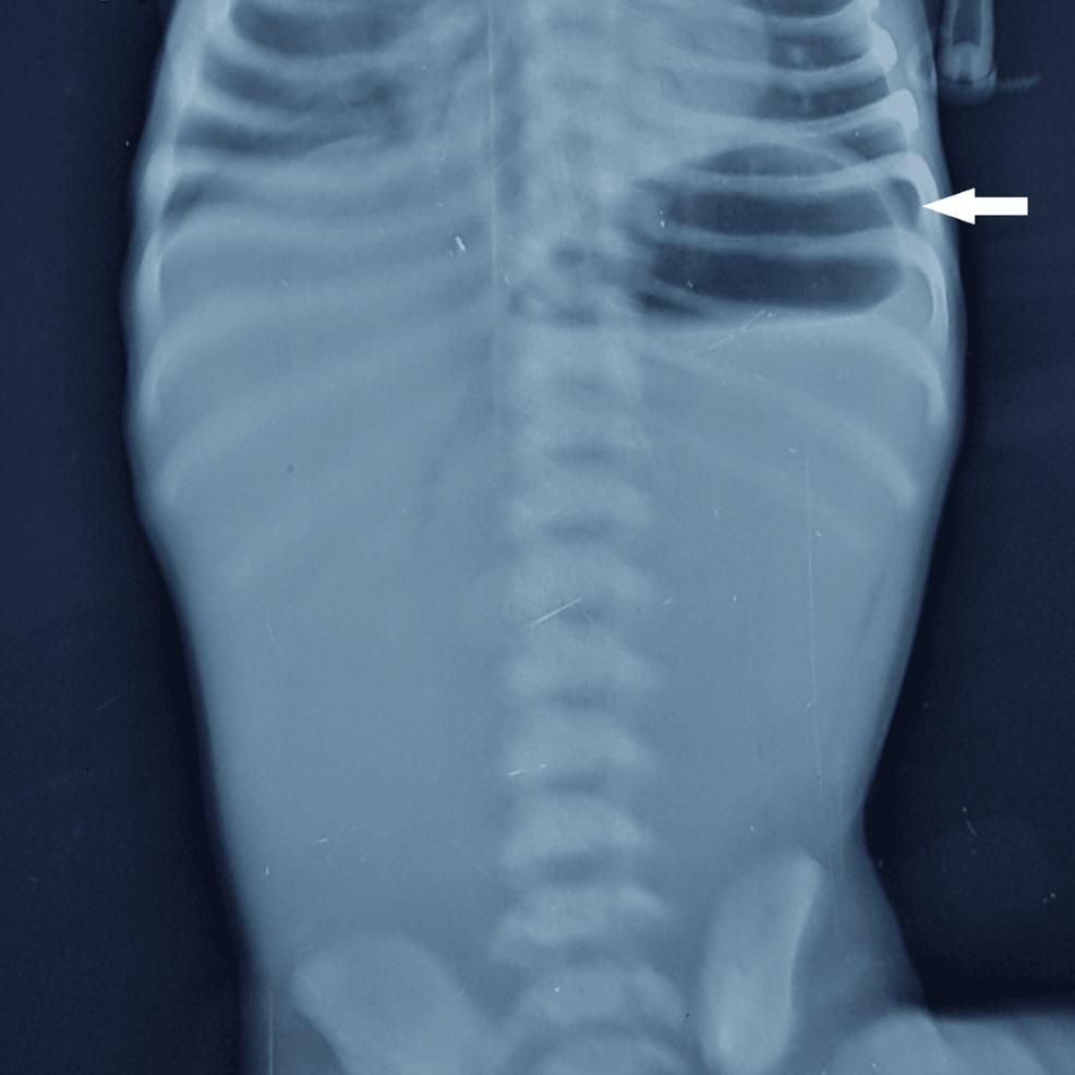
The infant arrived at the hospital with persistent non-bilious vomiting, lethargy, and signs of dehydration. Diagnostic imaging, including abdominal X-rays and ultrasound, confirmed the presence of a type 1 CPA—a condition where a mucosal diaphragm obstructs the stomach outlet.
Surgeons Repair Pyloric Diaphragm Before Infant Develops Bullae
On his seventh day of life, the neonate underwent an exploratory laparotomy. Surgeons successfully excised the pyloric mucosal diaphragm and performed a two-layer Heineke-Mikulicz pyloroplasty to restore gastrointestinal flow. The procedure was physically successful, but the case took an unexpected turn on the eighth postoperative day. The infant began to develop tense, fluid-filled bullae and superficial erosions on friction-prone areas like his elbows and feet.
This development, documented in Cureus, led to a clinical diagnosis of epidermolysis bullosa, a group of disorders characterized by extreme skin fragility. Unlike the severe, often fatal junctional EB—historically referred to as Carmi syndrome—this infant exhibited a milder, transient course. The lesions resolved spontaneously within two weeks, a pattern consistent with a self-improving dystrophic epidermolysis bullosa (DEB) phenotype.
Data from the National Center for Biotechnology Information (NCBI) shows the rarity of this condition: CPA accounts for less than 1% of all congenital gastrointestinal atresias, with an estimated prevalence of one in 100,000 live births. Clinical outcomes vary significantly depending on the underlying EB subtype.
| Feature | Classic Carmi Syndrome (JEB) | Self-Improving DEB Phenotype |
|---|---|---|
| Pathology | Cleavage within the lamina lucida | Anchoring fibril defects (COL7A1) |
| Prognosis | Historically poor; high mortality | Benign; spontaneous resolution |
| Long-term Scarring | Severe dystrophic scarring | Minimal or no scarring/milia |
Other reports, including a study archived by the National Library of Medicine, show that specialized wound management is essential for these infants. In one case, clinicians utilized hydrocolloid dressings and avoided adhesive tapes entirely to protect fragile skin during the healing process. These protocols, combined with aggressive fluid resuscitation and nutritional support, have become the standard for improving survival rates in neonates who previously faced a 50% to 70% mortality risk. A child was discharged on the 13th day when significant epithelialization in 90% of the lesions had occurred, highlighting the importance of meticulous care.
Financial Constraints Prevent Definitive Genetic Testing
Despite the successful outcome in this case, the path to a definitive diagnosis was hampered by financial constraints. While the medical team recommended a skin biopsy for immunofluorescence mapping and a multigene panel to confirm the EB subtype, the parents were unable to pursue these tests. As a result, the specific genetic defect remains a presumptive clinical diagnosis.
By age two, the child in the Cureus report was thriving, with only minor residual skin patches and no signs of the scarring typically associated with more severe blistering disorders. However, not all cases share this positive outcome. A case study from Source 3 describes a female baby who, despite initial stabilization, developed severe sepsis with multiorgan dysfunction and expired on the 25th day of life, underscoring the critical need for early intervention and resource availability.
As medical teams continue to refine their approach to these rare syndromic presentations, the focus remains on balancing life-saving surgery with the delicate, long-term care required for skin integrity. This patient's survival shows that even when a diagnosis historically has a poor prognosis, modern neonatology protocols can lead to recovery.
Preoperative Laboratory Findings
Preoperative laboratory investigations revealed abnormal values in key parameters. C-reactive protein was 0.71 mg/dL (0.02–0.30 mg/dL). These findings highlight the systemic stress and potential complications faced by the neonate.