Acute Hemobilia Following Liver Biopsy: A Rare Medical Emergency
Percutaneous liver biopsy has long been a foundational tool for diagnosing hepatic lesions, yet every invasive procedure carries an inherent risk. While clinicians often focus on the standard monitoring protocols for post-procedural recovery, a rare case report published in Cureus serves as a stark reminder that life-threatening complications can manifest with alarming speed. A 55-year-old man, undergoing a routine ultrasound-guided biopsy for a LI-RADS 4 hepatic lesion, experienced acute symptomatic hemobilia just 10 minutes after the procedure—a stark departure from the typical presentation window of several days.
The Rapid Onset of Symptoms
The patient, who had a history of chronic hepatitis B, was referred for a biopsy to evaluate a 40×39 mm liver lesion. The procedure itself, which utilized a 16G semi-automatic core biopsy needle, proceeded without immediate technical complications. However, the situation shifted rapidly when the patient developed sudden, intense right upper quadrant pain followed by hematemesis shortly after the biopsy was completed.
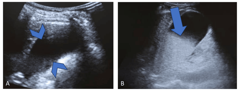
Bedside ultrasound provided the first critical clue, revealing echogenic material filling approximately half of the gallbladder lumen. This was a dramatic change from the clear, anechoic gallbladder observed just moments before. Because most post-biopsy complications develop over days, this rapid presentation requires immediate diagnostic vigilance in the recovery room.
The Diagnostic Role of Imaging
Following the ultrasound, the medical team utilized multiphasic computed tomography (CT) to better understand the internal landscape. The imaging confirmed hyperattenuating material within the gallbladder, consistent with acute blood products. While the CT did not identify an active site of contrast extravasation—meaning the specific vascular-biliary communication could not be pinpointed—the clinical context was clear. According to the data reported in Cureus, the absence of an identifiable vascular lesion on initial imaging does not rule out hemobilia, as the bleeding may be intermittent or self-limiting.
The patient’s case was managed conservatively, as he remained hemodynamically stable. His hemoglobin levels remained steady at 12.3 g/dL, and he was discharged after three days of observation. This successful outcome shows the value of a multidisciplinary approach to rare hemorrhagic events, where stable vital signs justified avoiding aggressive interventions such as angiography or embolization.
Contextualizing Hemorrhagic Complications
Hemobilia remains one of the rarest hemorrhagic complications of liver biopsies. Research indicates an incidence rate of approximately 0.06 per thousand procedures, based on a massive retrospective study of over 68,000 cases. Because it is so infrequent, the medical community lacks a standardized “best practice” for every scenario, forcing radiologists and surgeons to rely on sequential imaging—combining the real-time insights of ultrasound with the structural detail of CT—to guide their clinical judgment.
The “Quincke triad”—the classic clinical presentation of hemobilia consisting of gastrointestinal bleeding, right upper quadrant pain, and jaundice—is rarely seen in its entirety. This patient exhibited only two of those symptoms, emphasizing that physicians must maintain a high index of suspicion even when the full, textbook clinical picture is absent. For those in the medical field, this case emphasizes that the “immediate post-biopsy period” is not merely a waiting game; it is a critical window for active observation.
The message for patients is clear: while percutaneous biopsies are generally safe and remain the gold standard for tissue confirmation, they carry risks. For patients with LI-RADS 4 lesions, the benefit of obtaining a definitive diagnosis must always be weighed against the potential for rare but serious vascular injuries. As imaging technology becomes more sophisticated, the ability to catch these complications in the first 10 minutes—rather than the first five days—may define the difference between a routine recovery and a medical crisis.