Researchers at Washington University School of Medicine in St. Louis have discovered that the loss of the maternal UBE3A gene in Angelman syndrome causes an abnormal buildup of asparagine synthetase, tricking developing neurons into a false emergency stress mode that halts protein synthesis and stunts brain growth.
Uncovering the Molecular Culprit in UBE3A Loss
Angelman syndrome is a severe neurodevelopmental disorder driven by the loss of UBE3A, a gene that encodes an E3 ubiquitin ligase. This housekeeping protein tags surplus or unneeded proteins for cellular destruction across the brain.
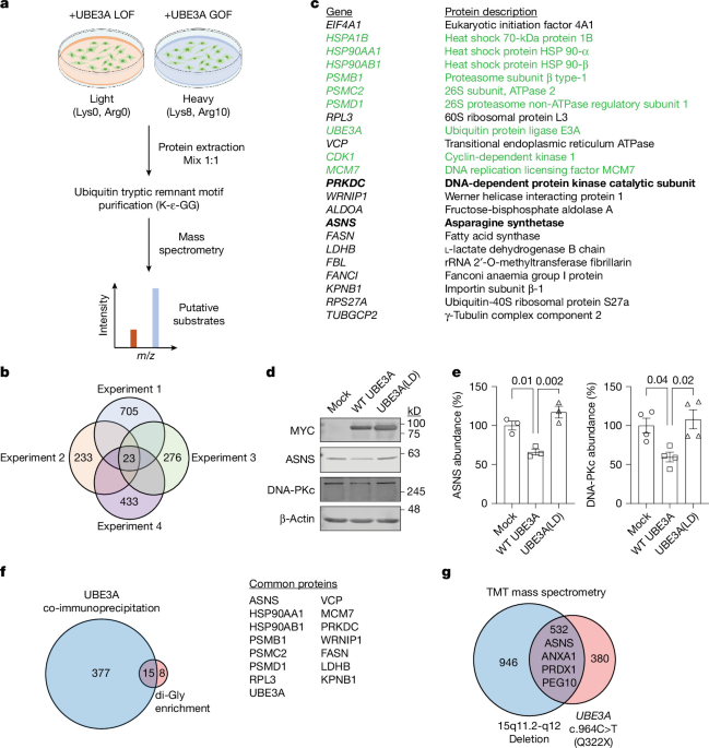
To isolate the driving mechanism, a research team led by senior author Jason Yi, assistant professor of neuroscience at WashU Medicine, generated cell models lacking functional UBE3A. Through systematic proteomic screening using SILAC and LC–MS/MS, the team identified 23 candidate proteins that abnormally accumulated in the gene’s absence. Among these candidates, one metabolic enzyme stood out: asparagine synthetase, or ASNS.
How Asparagine Synthetase Triggers False Neuronal Stress
Under ordinary physiological conditions, asparagine synthetase is harmless. However, in genetically modified mouse models and human neurons derived from patient cell lines, the uninhibited accumulation of ASNS activates a persistent cellular stress response within the brain. This buildup does not engage canonical cellular stress sensors or cause direct physical damage to cellular membranes. Instead, its excess serves as an aberrant biochemical trigger that signals to the cell that resources are depleted.
The work is detailed in a study titled Persistent neuronal stress signaling underlies Angelman syndrome phenotypes,
authored by Kwang-Soo Kim, Cory Gardner, Alissa Cullen, Jalin A. Stelzer, Kevin Cho, Ashish Sharma, Mati T. Nemera, Travis Law, Huma Naz, Guoyan Zhao, Harrison W. Gabel, Gary J. Patti, Erik S. Musiek, Tae-Hyuk Ahn, and Jason J. Yi.
The study utilized mice maintained on a C57BL6/J background. Angelman mice (Ube3am−/p+) were generated by crossing Ube3am+/p− heterozygous females with wild-type males. Utilizing a guide RNA, the Washington University Mouse Genetics Core produced an Asns-heterozygous mouse featuring a single-nucleotide deletion at the Q17 codon on exon 2 of Asns (chr. 6: 7689452, GRCm39 assembly). All experimental work involving animals received approval from the Institutional Animal Care and Use Committee (IACUC) at Washington University in St. Louis and was conducted in strict compliance with its established guidelines. Housing for the animals was provided in a specific-pathogen-free environment overseen by the Division of Comparative Medicine at Washington University School of Medicine, an institution holding full accreditation from AAALAC International.
For cellular assays described in the work, cells were maintained in Dulbecco’s modified Eagle’s medium pre-supplemented with GlutaMAX, fetal bovine serum, and an antibiotic–antimycotic solution containing penicillin, streptomycin, and amphotericin B. Primary cortical neurons were cultured and maintained using the B-27 Plus neuronal culture system supplemented with N2 and penicillin–streptomycin, with half of the culture medium replaced every two days until the desired day of the experiment. For proteasome inhibition assays, cells were treated with 15 μM MG132 for 12 hours before collection, while cells treated with an equal volume of dimethyl sulfoxide served as vehicle controls. Lysates were pre-cleared using protein G Sepharose beads for one hour at 4 °C on an orbital shaker, and protein bands were analyzed using an Odyssey CLx infrared imaging system and Image Studio software.

The neurons are tricked into thinking that they’re in stress when they’re not — that’s bad because they will then not grow normally.
Dr. Yi, assistant professor of neuroscience at WashU Medicine, via Neuroscience News
This unwarranted defense mechanism forces developing neurons to conserve cellular resources by dramatically curtailing protein synthesis. The observation that monosomes remain relatively unchanged while polysomes fall points to a selective defect in translation elongation or initiation rather than a global collapse of ribosome abundance.
Genetic Correction and Behavioral Rescue in Mouse Models
To test whether ASNS overabundance actively contributes to pathology rather than acting merely as a marker, the investigators introduced an Asns nonsense mutation into the Angelman mouse background. Genetic correction of the protein overabundance restored neuronal protein synthesis, corrected microcephaly, and improved many behavioral deficits, including open-field center time, hindlimb clasping, and marble burying.
At the same time, the rescue was partial rather than complete. Sleep cycles were not rescued, showing that UBE3A likely regulates multiple pathways alongside ASNS.
Implications for Future Therapeutic Interventions
The study, published in Nature, reframes Angelman syndrome as a disorder of persistent neuronal stress and impaired translational homeostasis. Nevertheless, the findings establish ASNS and the integrated stress response as viable targets that could complement genetically targeted approaches in future therapeutic development.
We haven’t been able to identify all of the things that the missing gene is doing and rescue the things that it is not doing in Angelman’s syndrome. But I think our data are the most complete so far to begin answering those questions.
Dr. Yi, via Neuroscience News
Readers should consult qualified medical professionals regarding neurodevelopmental conditions, as these research findings do not constitute direct medical advice or a validated clinical protocol. These insights provide a crucial foundation for future research aimed at translating these stress response mechanisms into effective treatments for individuals affected by the condition.


